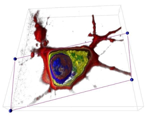
Nanolive SA, a start-up company founded in November 2013 at the EPFL Innovation Park in Lausanne, Switzerland, has developed a revolutionary microscope which allows for the very first time the exploration of a living cell in 3D without damaging it.
Since the discovery of the cell as the basic unit of life in the 17th century, scientists all around the world have been trying to investigate it deeper and deeper. This was not an easy task since they had to face two major problems: the cell is very small (10–6 meters) and it is transparent. The only way to solve these issues was to increase the complexity of the microscopes in order to gain resolution and to stain the cells in order to make them visible by adding chemical reagent or modifying them genetically.
This was still the case until today, even with the latest Nobel Prize technologies: the 2014 Nobel Prize for chemistry was assigned to S. Hell, E. Betzig, and W. Moerner who made revolutionary discoveries in the resolution of fluorescent microscopy.
Nanolive completely opposes this trend and, as a result, Nanolive’s microscope offers unperturbed and earlier unmet insights into the living cell: no need for any special procedures, which require intensive and long preparation. As no chemistry or marker is used at all, the observation is completely non-invasive to the cell, and allows resolving the cell’s parts down to 200nm.
“You really need to be able to look at living cells because life is animate – it‘s what defines life,“ Eric Betzig stated in a recent interview.
The 3D Cell Explorer responds to this desire by displaying the cell in a completely new way with a comprehensive representation of its morphology. Since the cell is the basis of all life on earth, this is a major milestone in the history of microscopy, which may change all the rules in the fields of Education, Biology, Pharmaceutics, Cosmetics, Labs and Industry.
The 3D Cell Explorer is based on an enabling technology that overcomes the limitation of light. Similar to a MRI/CT-scan in hospitals for the human body, our product makes a complete tomography of the refractive index within the living cell. For the first time you can actually look inside the cell and discover its interior such as nucleus and organelles. Thanks to the 3D Cell Explorer never again researchers will have to “guess” what happens inside a living cell. They will actually see and measure precisely the impact of stimuli and drugs on cells, thus enabling completely new fields of research and smarter products.
Nanolive’s technology detects the physical refractive index of the different cell parts with resolution far beyond the diffraction limit. Nanolive’s microscope, the 3D Cell Explorer, can be handled without special training and is sold worldwide at an affordable price (19’900 €).
For the same purpose the company has developed an intuitive and unique software called STEVE. To mark and label certain parts of the measured cells in 3D, the software allows the user to “virtually brush” and “digitally paint” the part of a cell based on their physical properties (called refractive index). STEVE will automatically detect all regions with same refractive index characteristics (different organelles have different optical properties) and digitally stain them with the same color. This process is quantitative and can be applied for a limitless amount of colors. Changes to digital stains are shown in real time in both 2D slices and 3D view. Stains panels can be constantly modified by the user, saved and reused for other cells of the same line.
A trial version of STEVE is directly downloadable from the company’s website. For additional support, Nanolive provides a getting started video and getting started document.
The 3D Cell Explorer is a tool for discovery and we are just at the beginning of exploring all the potential fields of research. It allows the measurement of cellular processes and kinetics in real-time enabling multi parameter analysis at single-cell and sub-cellular scales. Some of the fields of applications for the Nanolive technology are:
- Cell division
- Cell morphology monitoring
- Cell differentiation (200+ types)
- Cell-cell interaction
- Intracellular trafficking
- Cellular remodeling processes
- Cell death (apoptosis, autophagy, necrosis)
- Viral/ bacterial inclusions
- Cell proliferation
- Drug monitoring (i.e. cancer drugs)
- In vitro fertilization
- Nanoparticle internalization
Help keep news FREE for our readers
Supporting your local community newspaper/online news outlet is crucial now more than ever. If you believe in independent journalism, then consider making a valuable contribution by making a one-time or monthly donation. We operate in rural areas where providing unbiased news can be challenging. Read More About Supporting The West Wales Chronicle


